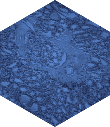
FOLLICULAR LYMPHOMA AND MARGINAL ZONE LYMPHOMA (FL/MZL)

FL and MZL are two forms of indolent (slow growing) non-Hodgkin’s lymphoma (NHL)—the 8th leading cause of US cancer deaths in 2020.1,2 Both FL and MZL are generally considered to be chronic and incurable diseases characterized by multiple recurrences and relapses.3,4
FL is the most common form of indolent NHL and accounts for ~17.1% of all NHL cases.5
More than 13,000 new cases of FL are expected in the US in 2021, with ~12,500 patients with relapsed/refractory (R/R) FL on therapy each year.6,7
MZL is the second most common form of indolent NHL and accounts for ~10.6% of all NHL cases.5 More than 8000 new cases of MZL are expected in the US in 2021, with ~6000 patients with R/R MZL on therapy each year.6,7
Number of new cases and percent of total NHL cases
FL

13,200
MZL

8,200
FL AND MZL PATHOPHYSIOLOGY
FL and MZL are malignancies that arise from B cells.1 B cells originate in the bone marrow and move into the lymphoid tissue, where they mature into effector cells, and ultimately memory B cells or plasma cells.8 Chromosomal translocations and/or mutations can arise during B-cell development within the lymphoid follicle, and these may contribute to the development of either FL or MZL.8,9

Mutations that occur during B-cell development are common, however most of these pre-cancerous cells are destroyed by the immune system.8,11 However, a dysfunctional immune system may not be able to detect malignant (or neoplastic) cells, which can lead to the development of lymphoma.12 Patients with weakened or dysfunctional immune systems (i.e. suffering from infections or autoimmune diseases) are at greater risk of developing lymphoma.10
Because FL and MZL arise from the uncontrolled proliferation of B cells, the BCR signaling pathway plays an important role in driving the proliferation and survival of lymphoma cells.14 Activation of the BCR results in the activation of downstream kinases, such as spleen tyrosine kinase (SYK), bruton tyrosine kinase (BTK), and phosphoinositide-3 kinase (PI3K, specifically PI3K-delta—an isoform that is highly expressed in B cells).14
SYK
SYK plays a role in lymphoma cell adhesion.15
BTK
BTK is important for cell proliferation, differentiation, and allows lymphoma cells to communicate with the immune microenvironment.14,15
PI3K-delta
PI3K-delta promotes cell proliferation, survival, and migration of malignant B cells.15

PI3K proteins comprise a family of enzymes that can exist as 1 of 4 different isoforms (α, β, δ, γ) and regulate many signaling pathways involved in cell proliferation and survival.19 PI3Kδ is a key mediator of BCR signaling.19 Downstream of PI3Kδ are the proteins, AKT and mTOR, so this pathway is often referred to as the PI3K-AKT-mTOR pathway.19 Given the importance of the PI3K pathway in cancer, it is critical to understand the role of each PI3K isoform and the different cell types that are affected.
- PI3Kα
- PI3Kβ
- PI3Kδ
- PI3Kγ
- Tissue/cell expression20-24
-
- Broad distribution
-
- Broad distribution
-
- B cells
-
- T cells, macrophages neutrophils
- Primary Function20,23-27
-
- Plays a role in tumorigenesis
- Involved in insulin signaling and glucose metabolism
-
- Involved in platelet activation
- Plays a role in the development to thrombotic diseases
-
- Involved in the activation, proliferation, and survival of B cells as well as antibody production
-
- Involved in the innate immune response, suppress inflammation
- Biological response when inhibited19,20,22,25
-
- Blocks glucose uptake, resulting in hyperglycemia
-
- Defects in thrombosis, due to its effects on platelets
-
- Enhance anti-tumor immunity
-
- Activate T cells and macrophages to produce an inflammatory and/or autoimmune-like response
Tissue/cell expression20-24
Broad distribution
Primary function20,23-27
Plays a role in tumorigenesis
Involved in insulin signaling and glucose metabolism
Biological response
when inhibited19,20,22,25
Blocks glucose uptake, resulting in hyperglycemia
Tissue/cell expression20-24
Broad distribution
Primary function20,23-27
Involved in platelet activation
Plays a role in the development to thrombotic diseases
Biological response
when inhibited19,20,22,25
Defects in thrombosis, due to its effects on platelets
Tissue/cell expression20-24
B cells
Primary function20,23-27
Involved in the activation, proliferation, and survival of B cells as well as antibody production
Biological response
when inhibited19,20,22,25
Enhance anti-tumor immunity
Tissue/cell expression20-24
T cells, macrophages neutrophils
Primary function20,23-27
Involved in the innate immune response, suppress inflammation
Biological response
when inhibited19,20,22,25
Activate T cells and macrophages to produce an inflammatory and/or autoimmune-like response
CLINICAL RESEARCH IN LYMPHOMA
We are researching new combinations and other targeted agents for B-cell malignancies.

TG Therapeutics is committed to researching
multiple targets in NHL.
References: 1. Lymphoma Research Foundation. Understanding Non-Hodgkin Lymphoma: A guide for patients, survivors, and loved ones. https://lymphoma.org/wp-content/uploads/2021/04/LRF-NHL-Booklet_4.21.pdf. Accessed November 15, 2021. 2. National Cancer Institute. SEER Cancer Stat Facts: Common Cancer Sites. https://seer.cancer.gov/statfacts/html/common.html. Accessed January 14, 2021. 3. Rivas-Delgado A, et al. Brit J Haematol. 2019;184(5):753-759. 4. Denlinger NM, et al. Cancer Manag Res. 2018;10:615-624. 5. National Cancer Institute. SEER Cancer Statistics Review 2008-2017: Non-Hodgkin Lymphoma. Table 19.26. https://seer.cancer.gov/csr/1975_2017/results_single/sect_19_table.26_2pgs.pdf. Accessed October 14, 2021. 6. National Cancer Institute. SEER Cancer Stat Facts: Non-Hodgkin Lymphoma. https://seer.cancer.gov/statfacts/html/nhl.html. Accessed October 14, 2021. 7. TG Therapeutics. Data on file. 8. Küppers R. Nat Rev Cancer. 2005;5(4):251-262. 9. Huet S, et al. Nat Rev Cancer. 2018;18(4):224-239. 10. American Cancer Society. Non-Hodgkin Lymphoma Risk Factors. https://www.cancer.org/cancer/non-hodgkin-lymphoma/causes-risks-prevention/risk-factors.html. Accessed October 14, 2021. 11. Swann JB, Smyth MJ. J Clin Invest. 2007;117(5):1137-1146. 12. Kridel R, et al. J Clin Invest. 2012;122(10):3424-3431. 13. Dong S, et al. J Clin Invest. 2019;129(1):122-136. 14. Burger JA. N Engl J Med. 2020;383(5):460-473. 15. Burger JA, et al. Trends Immunol. 2013;34(12):592-601. 16. MacDonald D, et al. Curr Oncol. 2016;23(6):407-417. 17. Sindel A, et al. Blood. 2018;132 (1 suppl):4125. 18. Dienstmann R, et al. Mol Cancer Ther. 2014;13(5):1021-1031. 19. Fruman DA, et al. Cell Reports. 2017;170(4):605-635. 20. Kaneda MM, et al. Nature. 2016;539(7629): 437-442. 21. Ali AY, et al. Leukemia. 2018;32(9):1958-1969. 22. Ali K, et al. Nature. 2014;510(7505):407-411. 23. Esposito A, et al. JAMA Oncol. 2019;5(9):1347-1354. 24. Ortiz-Maldonado V, et al. Ther Adv Hematol. 2015;6(1):25-36. 25. Nylander S, et al. J Thromb Haemost. 2012;10(10):2127-2136. 26. Curigliano G, et al. Drug Saf. 2019;42(2):247-262. 27. Durand CA, et al. J Immunol. 2009;183(9):5673-5684.

